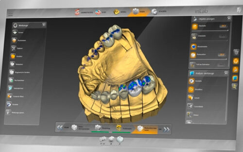
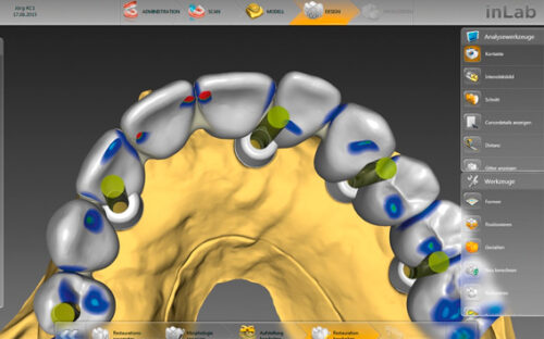
Custom abutments, screw-retained bridges and bars, and surgical guides. The implantology module of inLab SW 16.0 includes all the CAD tools and features that are necessary for customised restorations on single and multiple implants, as well as for the design of complete surgical guides.
Features include:
- New: Implant-level screw retained bridges and bars
- Bridges and screw retained bars in multi-unit abutments
- Customised abutments (zirconium or titanium)
- Surgical guides (integrated implantology)
Follow the real movement on the move
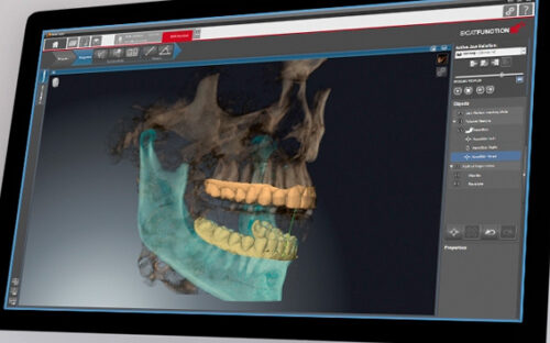
Sicat Function is the first integrated digital 3D solution that visualises the actual movement of the individual patient's lower jaw within the 3D volume. Anatomical traces of the temporomandibular joint can be displayed for every possible position of the volume.
- Direct visualisation of the anatomically correct movement of the mandible, including the actual condyle-fossa relationship
Diagnosis, planning and consultation with your patient in a single session and without leaving your clinic
Integration with CEREC for a digital 100 % workflow
The new TAT (temporomandibular joint disorder) software for every clinic.
Direct visualisation of anatomically correct jaw movement
Actual condyle-facial relationship during jaw movement, anatomically correct trajectory, specific positioning of the trajectory in the 3D volume (if necessary, also in comparison to conventionally used axial points), evaluation of the occlusion based on integrated optical surface studies.
All in one session and without leaving the clinic
Diagnosis, planning and consultation with the patient.
https://www.youtube.com/watch?v=rDbab8MDWN4
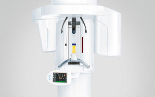
Orthophos SL 3D
The true multifunctional imaging unit
With Orthophos SL, Clínica Dental Cots is prepared for a wide range of treatment situations. On the 2D side, the revolutionary DCS sensor and SL technology meet the requirements of clinicians with very high demands for panoramic images. In 3D, you can choose between the 8 cm x 8 cm or 11 cm x 10 cm volume unit, both of which offer different collimations.
Patients appreciate the soothing ambient light during the imaging process.
Revolutionary 2D DCS imaging for efficient diagnosis
Thanks to DCS (Direct Conversion Sensor) technology that skips light conversion, it retains maximum image information, benefiting from superb image sharpness.
Sharp Layer technology automatically adapts the panoramic curve to the patient's individual anatomical features, ensuring that the entire jaw is always in the image.
More volumes for more treatment options.
A selectable field of view starting with a focused volume of 5 cm x 5.5 cm and going up to 11 cm x 10 cm for upper airway analysis allows you to offer a wider range of treatments in your clinic.
Automatic assistance for patient positioning, optimising the session.
The new patented occlusal bite block, the Easy Volume Indicator lights and the 3-point head fixation: perfect positioning for correct diagnosis and scanning has never been easier.
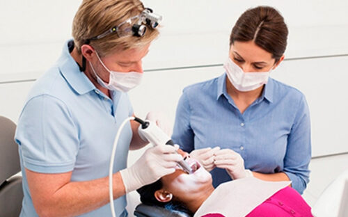
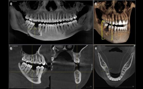
3D technology is becoming more and more established in dental clinics around the world. For more precise diagnoses and treatment explanations, 3D X-rays offer several advantages. Whether for overlapping teeth, unexpected runs of nerve canals, hidden roots or abnormalities of the temporomandibular joints, 3D images are invaluable for a large number of diagnoses.
Orthophos SL 3D offers a wide range of options to meet these needs: different volume sizes to choose from, HD/SD/Low radiation dose modes and intelligent, intuitive software to make the most of the images obtained and link them directly to treatment.